Archive

Tag Archives: medical illustration

After final presentations a couple of days ago, school has let out for the semester and I have been highly anticipating this time to relax a bit before summer semester begins. Looking back on the last few months, I feel very proud of my program, my classmates, and the work that we’ve all made this semester. Additionally, it’s been really fun to teach BIOS223 (Cell Biology Laboratory) and learning new things along the way. Even though Neuroanatomy ended halfway through the semester, I’m so happy that I took that class. Not only did I learn a ton about the brain, but I also got to study and attend lab with my classmate Jill Tessler. It was a great experience to share with another BVIS student!

To finish out the semester, we worked on a project for Illustration Techniques that was a little out of the ordinary. We had to complete an editorial illustration. This took a lot of patience, trial and error, but it finally worked out in the end. Additionally, I completed another editorial illustration for the same journal that our illustrations from class will be submitted to — the Northwestern University Public Health Review.

Image


An illustration for the Spring edition of NPHR for an article on sleep deprivation and its effect on the brain.

Image

This illustration is for an article on energy and its role in public health. It will be submitted for publication in the Fall 2014 issue of NPHR.

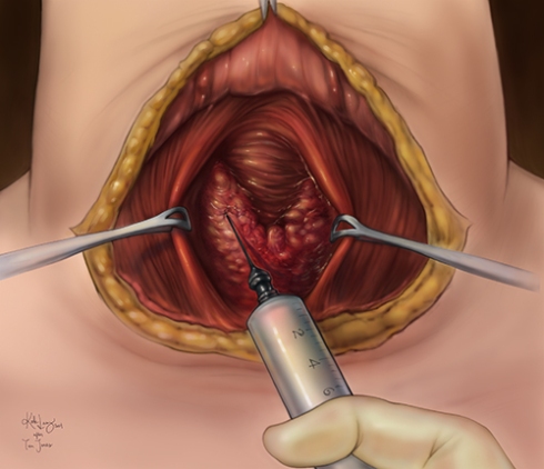
Before this final assignment, we did a Tom Jones reproduction piece for Illustration Techniques. Taking a black and white watercolor illustration done by the founder of the BVIS program, Tom Jones, we were asked to add color using Photoshop. I chose an illustration of a thyroid surgery. This was the final result.

Image

A digital, color reproduction painting of a Tom Jones illustration.

After all that, I’m ready to take a break to read some novels, do some sketching, and enjoy the nice weather (finally!).

This semester, so far, has been a real mix of classes and assignments. The first eight weeks were dominated by the Neuroanatomy course, while the second eight were spent practicing and solidifying skills and concepts presented in my studio courses. In addition, I started my first teaching assistantship. It has been a wonderful opportunity to get a view inside the different learning styles of undergraduate students while also re-learning the concepts of Cell Biology (and not to mention learning how to operate a Zeiss fluorescence microscope, wow!!). I’ve also just begun using the I-share library system, which I’m sure will come in handy once my research begins to take off. So far I’ve checked out a few books on developmental biology and grammar. After grading seemingly endless lab reports this semester, I’ve gained some interest in correct grammar and writing technicalities to ensure my writing can communicate as well as possible. With the end of the semester just around the corner, I only have a few projects and a final presentation to complete, and then there will be some time to recuperate from my first year in the BVIS program. It’s been amazing so far, and I’m beyond excited to see what my class comes up with this summer!

As per usual, here are a few images of what I’ve been working on lately.

Image

Exercise 8 for Computer Visualization

This exercise helped the class learn the power of the program Mimics, which allows a user to input medical imaging data (like MRI) and construct a 3D model from that data. The mandible that you see in this image was taken from the Visible Human Project male dataset. My classmates and I used Mimics to scroll through the individual MRI slices and mask out the mandible from the rest of the tissue. We had to do this for each slice, but the program has a couple of options that make this process somewhat less taxing. After we masked out the data, we saved a file that could be imported into Max. This model has an excess of polygons that make the model’s functionality less than optimal, so to alleviate this issue we re-topologized the model by drawing on our own polygons. Eventually, the model was brought into Mudbox for some painting and this was the result.

Our last exercise for Illustration Techniques was to digitally reproduce part of a carbon-dust drawing from one of the masters of medical illustration, Max Brödel.

Image

The original Brödel drawing from 1917.

Max Brödel was one of the pioneers of modern medical illustration, and started the first program at Johns Hopkins University in the early 20th century. He was a master of anatomical drawing, pen and ink, and carbon-dust techniques. His work is delicate, nuanced, extremely accurate, and beautiful.

brodel-Lamysm

…and my digital reproduction.

At first, I felt afraid of butchering his wonderful work by reproducing it myself, but in the end I learned so much from this exercise. Brödel uses tone with such success, and by studying his technique I feel I am a lot more at ease with digital rendering.

Next, I’ll be re-topologizing kidneys and doing another digital reproduction piece, this time originally by Tom Jones, the founder of the BVIS program! I’m really excited, as this piece is all about color (finally!).